Osteology
Learning Objectives
- Define osteology and explain why bone structure matters in anatomy, pathology, and surgery.
- Classify bones by shape and relate each category to common examples in the human body.
- Describe the gross and microscopic structure of bone, including cortex, trabeculae, periosteum, and marrow.
- Explain endochondral and intramembranous ossification and connect them to normal growth and fracture healing.
- Identify common bone markings and explain why they matter for muscle attachment, joint movement, and neurovascular relations.
- Apply osteological concepts to fractures, osteoporosis, osteomyelitis, and forensic identification.
Quick Answer
Osteology is the study of bones: their structure, development, function, and clinical significance. Bones do much more than provide a rigid frame. They protect organs, act as levers for movement, store minerals such as calcium and phosphate, and contain marrow that produces blood cells. Medical students study osteology to understand fracture patterns, joint mechanics, skeletal development, surgical landmarks, and radiographic interpretation. If you know how a bone is built, how it grows, and what structures attach to it, you can reason through many clinical problems instead of memorizing them in isolation.
What Is Osteology?
Osteology is the branch of anatomy that focuses on the skeleton. It studies individual bones, their landmarks, their internal organization, and the way they interact with joints, muscles, nerves, and vessels.
In practice, osteology helps answer questions such as:
- Why does a fracture at one part of a bone heal differently from a fracture at another part?
- Why are some bony prominences easy to palpate and useful as surface landmarks?
- Why does damage near the surgical neck of the humerus threaten the axillary nerve?
- Why do children develop different fracture patterns from adults?
This is why osteology sits at the intersection of anatomy, orthopedics, radiology, pathology, and forensic medicine.
Why Bones Matter Clinically
Bones are living tissues, not passive supports. They matter clinically because they:
- Protect vital organs: the skull protects the brain, ribs protect thoracic organs, and vertebrae protect the spinal cord.
- Enable movement by serving as levers for muscles across joints.
- Store minerals, especially calcium and phosphate, which are essential for nerve conduction and muscle contraction.
- House bone marrow, the site of hematopoiesis in many bones.
- Preserve evidence of growth, trauma, infection, and nutritional disease.
For example, a neck of femur fracture is not just a broken bone. Its location affects blood supply to the femoral head, the risk of avascular necrosis, mobility outcomes, and the choice of surgical repair.
Bone Classification
Bones are commonly classified by shape because shape usually hints at function.
| Type of Bone | Main Features | Examples | Why It Matters |
|---|---|---|---|
| Long bones | Longer than wide; shaft with two ends | Femur, humerus, radius | Built for leverage and movement |
| Short bones | Roughly cube-shaped | Carpals, tarsals | Provide stability with limited motion |
| Flat bones | Thin, often curved plates | Skull vault, sternum, ribs, scapula | Protect organs and offer broad muscle attachment |
| Irregular bones | Complex shapes | Vertebrae, sphenoid, hip bone | Specialized functions and multiple attachments |
| Sesamoid bones | Develop within tendons | Patella | Improve mechanical advantage and reduce friction |
| Pneumatic bones | Contain air spaces | Frontal, ethmoid, maxilla | Reduce weight and contribute to resonance |
Example
The patella is a sesamoid bone embedded in the quadriceps tendon. It increases the leverage of the quadriceps across the knee, improving extension efficiency.
Real-World Example
Scaphoid fractures are common because the scaphoid is a small carpal bone that transmits force during falls on an outstretched hand. Its blood supply is vulnerable, so fracture location matters.
Why It Matters
Knowing the bone type helps predict function, blood supply, fracture behavior, and imaging appearance.
Common Misunderstanding
Students sometimes assume all bones are structurally similar and differ only in size. In reality, a vertebra, patella, and femur are built differently because they do different jobs.
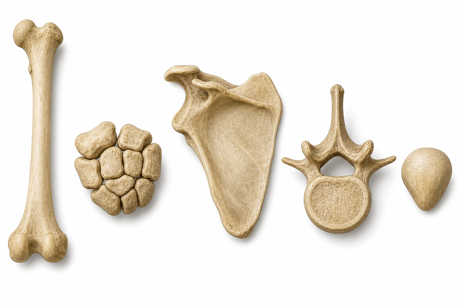
Figure 1. This comparison plate makes the shape-based classification easier to remember because each category is shown side by side rather than as a text-only list.
Gross Structure of a Typical Bone
A typical long bone illustrates most core osteological concepts:
- Diaphysis: the shaft, mainly compact bone surrounding a medullary cavity.
- Epiphysis: the expanded ends, rich in cancellous bone and articular surfaces.
- Metaphysis: the region between diaphysis and epiphysis, especially active during growth.
- Articular cartilage: hyaline cartilage covering joint surfaces.
- Periosteum: fibrous outer covering important for nourishment, repair, and tendon attachment.
- Endosteum: inner lining of marrow spaces and medullary cavity.
- Medullary cavity: central cavity containing marrow.
Real-World Example
In an elderly patient with osteoporosis, trabecular bone loss in the vertebral bodies can lead to compression fractures even after minor trauma.
Why It Matters
Fracture location, healing potential, and surgical fixation all depend on which part of the bone is involved.
Common Misunderstanding
The metaphysis is not just a vague transition zone. In growing children, it is biologically active and clinically important in growth plate injuries and hematogenous osteomyelitis.

Figure 2. Use the cutaway to connect bone vocabulary with actual structure: this is the image to revisit before fracture-healing, remodeling, and osteoporosis questions.
Microscopic Structure and Bone Cells
Bone is dynamic tissue made of cells and extracellular matrix.
Main Bone Cells
| Cell | Main Function | Clinical Relevance |
|---|---|---|
| Osteoblasts | Form new bone matrix | Active in growth and healing |
| Osteocytes | Maintain bone tissue | Sense mechanical stress |
| Osteoclasts | Resorb bone | Important in remodeling and osteoporosis |
Bone Matrix
- Organic component: mainly type I collagen, giving tensile strength.
- Inorganic component: hydroxyapatite crystals, giving compressive strength.
Compact vs Cancellous Bone
- Compact bone is dense and organized into osteons, making it strong for weight-bearing.
- Cancellous bone has trabeculae and marrow spaces, making it lighter and metabolically active.
Real-World Example
In osteogenesis imperfecta, defective collagen weakens the organic matrix, so bones fracture easily even if mineralization is present.
Why It Matters
Different diseases affect different parts of the matrix. That is why rickets, osteoporosis, and osteogenesis imperfecta do not look identical.
Common Misunderstanding
Students often equate “more calcium” with “stronger bone.” Bone strength depends on architecture, collagen quality, remodeling balance, and load distribution, not mineral alone.
Ossification and Growth
Bone develops through two major processes.
Intramembranous Ossification
- Bone forms directly from mesenchyme.
- Produces many flat bones of the skull and part of the clavicle.
Endochondral Ossification
- Bone replaces a cartilage model.
- Produces most long bones and much of the axial skeleton.
Growth Plate Basics
Long bones lengthen at the epiphyseal plate. This is why injuries to the growth plate in children can disturb future limb length or shape.
Real-World Example
A Salter-Harris fracture involves the growth plate and can impair normal development if not recognized and managed properly.
Why It Matters
Developmental mechanism explains why pediatric bone injuries differ from adult fractures and why some congenital skeletal disorders affect specific bones.
Common Misunderstanding
Ossification is not finished at birth. Many bones continue to grow, fuse, and remodel throughout childhood and adolescence.
Bone Markings and Landmarks
Bone surfaces contain named features that reflect function.
| Marking | Meaning | Example | Importance |
|---|---|---|---|
| Tubercle / tuberosity | Projection for attachment | Greater tubercle of humerus, tibial tuberosity | Muscle and ligament insertion |
| Trochanter | Large projection | Greater trochanter of femur | Powerful muscle attachment |
| Spine / crest | Ridge-like projection | Iliac crest, spine of scapula | Palpable landmark |
| Condyle | Rounded articular surface | Occipital condyle, femoral condyles | Joint movement |
| Foramen | Opening | Foramen magnum, mental foramen | Passage for nerves and vessels |
| Fossa | Depression | Olecranon fossa, mandibular fossa | Articulation or accommodation |
Real-World Example
The mental foramen is an important landmark in dentistry because the mental nerve emerges here, and local anesthesia must account for its position.
Why It Matters
Landmarks guide palpation, surgery, imaging, and nerve blocks.
Common Misunderstanding
These names are not trivia lists. They tell you what happens at that location: attachment, articulation, passage, or depression.
Bone Remodeling and Healing
Bone constantly remodels in response to load, hormones, and injury.
Remodeling
- Maintains structural integrity
- Adapts to stress
- Helps regulate mineral balance
Fracture Healing
- Hematoma formation
- Inflammatory response
- Soft callus formation
- Hard callus formation
- Remodeling
Real-World Example
A tibial shaft fracture in a smoker may heal slowly because vascular supply, mechanical stability, and tissue biology all influence repair.
Why It Matters
Healing is not automatic or identical in every patient. Age, nutrition, infection, blood supply, and fixation all matter.
Common Misunderstanding
Students often think X-ray union and functional recovery happen at the same time. They do not. A fracture may look healed radiographically before full strength and normal loading return.
Clinical Correlations
Osteoporosis
Reduced bone mass and altered architecture increase fracture risk, especially in vertebrae, hip, and distal radius.
Osteomyelitis
Bone infection often localizes in metaphyseal regions because of blood flow patterns.
Avascular Necrosis
Some fractures threaten bone viability by interrupting blood supply, such as femoral neck fractures.
Forensic and Anthropological Relevance
Bones preserve clues about age, sex, trauma, nutritional status, and chronic disease.
Key Terms
| Term | Definition | Related Concept |
|---|---|---|
| Osteology | Study of bones | Skeletal anatomy |
| Diaphysis | Shaft of a long bone | Compact bone |
| Epiphysis | End of a long bone | Joint surface, growth |
| Metaphysis | Region between shaft and epiphysis | Growth plate, osteomyelitis |
| Periosteum | Fibrous covering of bone | Repair, attachment |
| Endosteum | Inner lining of bone cavities | Remodeling |
| Cancellous bone | Spongy trabecular bone | Marrow, lightweight support |
| Compact bone | Dense cortical bone | Strength, load-bearing |
| Ossification | Formation of bone tissue | Growth, development |
| Sesamoid bone | Bone embedded in a tendon | Patella |
| Osteoblast | Bone-forming cell | Healing |
| Osteoclast | Bone-resorbing cell | Remodeling |
Common Mistakes
Misconception: Bones are inert structures whose main job is support.
Why it's wrong: Bone is metabolically active tissue that remodels, stores minerals, responds to hormones, and houses marrow.
Correct understanding: The skeleton is a living organ system involved in movement, hematopoiesis, mineral homeostasis, and structural protection.
Misconception: All fractures heal the same way if the broken ends are simply aligned.
Why it's wrong: Healing depends on blood supply, bone type, age, infection, fixation, and patient factors such as smoking or malnutrition.
Correct understanding: Fracture healing is a biological process that varies by location and patient context.
Misconception: Bone markings are just names to memorize for exams.
Why it's wrong: Most markings indicate attachment sites, joint surfaces, or pathways for neurovascular structures.
Correct understanding: Bone landmarks are functional clues that help explain movement, palpation findings, imaging, and surgical anatomy.
Comparison and Connections
| Feature | Long Bone | Flat Bone | Irregular Bone |
|---|---|---|---|
| Typical role | Leverage and movement | Protection and broad attachment | Specialized support and protection |
| Common example | Femur | Parietal bone | Vertebra |
| Main development | Mostly endochondral | Often intramembranous | Mostly endochondral with mixed patterns |
| Common clinical issue | Shaft fracture, growth plate injury | Skull fracture | Vertebral compression, canal compromise |
| Imaging emphasis | Alignment and cortex | Sutures and plates | Complex 3D relationships |
Practice Questions
Recall
Q1. Name the main categories of bones by shape.
Answer guidance: Long, short, flat, irregular, sesamoid, and sometimes pneumatic bones.
Q2. What is the difference between periosteum and endosteum?
Answer guidance: Periosteum covers the outer surface of bone; endosteum lines internal marrow spaces and the medullary cavity.
Understanding
Q3. Why are metaphyseal regions important in pediatric bone disease?
Answer guidance: They are highly vascular, close to the growth plate, and important in both growth disturbances and hematogenous osteomyelitis.
Q4. Explain why compact and cancellous bone have different structural advantages.
Answer guidance: Compact bone resists heavy load and bending; cancellous bone provides lighter support and contains marrow while distributing forces through trabeculae.
Application
Q5. A patient fractures the neck of the femur after a fall. Why is this injury more concerning than a simple cortical break elsewhere?
Answer guidance: It can disrupt blood supply to the femoral head and lead to avascular necrosis, especially in older adults.
Q6. A child sustains a fracture involving the growth plate. What long-term issue should clinicians watch for?
Answer guidance: Disturbed longitudinal growth, angular deformity, or limb-length discrepancy.
Analysis
Q7. Compare osteoporosis with osteomalacia in terms of what is wrong with the bone.
Answer guidance: Osteoporosis primarily reduces bone mass and architecture; osteomalacia impairs mineralization of osteoid.
Q8. Why is knowledge of bone markings essential for both anatomists and surgeons?
Answer guidance: Markings identify attachment sites, passages, and articular regions, helping with orientation, exposure, and avoidance of nearby structures.
FAQ
Q: Why do children get greenstick fractures more often than adults?
Children's bones are less brittle and have thicker periosteum, so bending injuries may cause incomplete fractures rather than full breaks.
Q: Why is the periosteum important in fracture healing?
It contains blood supply, osteogenic cells, and attachment points that contribute strongly to repair and callus formation.
Q: What is the easiest way to remember bone markings?
Group them by function: projections for attachment, surfaces for articulation, and openings for passage.
Q: Why are vertebral fractures common in osteoporosis?
Vertebral bodies contain abundant trabecular bone, which is especially vulnerable to osteoporotic loss.
Q: Is marrow the same in all bones?
No. Red marrow is more active in hematopoiesis, while yellow marrow contains more fat; their distribution changes with age.
Quick Revision
- Osteology is the study of bones, their structure, development, and clinical relevance.
- Bones protect organs, support movement, store minerals, and contain marrow.
- Long, short, flat, irregular, sesamoid, and pneumatic bones differ by shape and function.
- A long bone includes diaphysis, metaphysis, epiphysis, periosteum, and marrow cavity.
- Compact bone is dense and strong; cancellous bone is trabecular and metabolically active.
- Osteoblasts build bone, osteoclasts resorb it, and osteocytes maintain it.
- Intramembranous ossification forms many flat bones; endochondral ossification forms most long bones.
- Bone markings reveal attachment sites, articular surfaces, and neurovascular passages.
- Fracture healing progresses through hematoma, callus formation, and remodeling.
- Osteology underpins orthopedics, radiology, surgery, and forensic interpretation.
Related Topics
Prerequisites: Introduction to Human Anatomy, anatomical position, and basic tissue terminology.
Related Topics: Gross Anatomy of Upper Limb, Gross Anatomy of Lower Limb, Histology, Embryology.
Next Topics: Surface Anatomy for palpable landmarks, then regional anatomy pages to see how bones organize limbs, thorax, and skull.