Surface Anatomy
Learning Objectives
- Define surface anatomy and explain how it connects external landmarks to deeper structures.
- Identify major bony, muscular, vascular, and visceral landmarks used in bedside examination.
- Use surface anatomy to localize pulse points, organ borders, and safe procedural areas.
- Relate visible or palpable findings to likely underlying structures and common pathologies.
- Distinguish reliable landmarks from variable features that can change with body habitus, age, or disease.
- Apply surface anatomy to clinical examination, injections, cannulation, and emergency assessment.
Quick Answer
Surface anatomy is the study of structures that can be seen, palpated, or projected onto the body surface. It is what turns anatomy from an abstract map into a bedside skill. When a clinician feels the apex beat, checks the radial pulse, estimates liver enlargement, marks an intercostal space for needle insertion, or avoids the sciatic nerve during gluteal injection, they are using surface anatomy. It matters because patients are examined from the outside, while the structures we need to assess are deeper. Surface anatomy bridges that gap.
What Is Surface Anatomy?
Surface anatomy focuses on external landmarks that help identify deeper bones, muscles, vessels, nerves, organs, and spaces. It relies on inspection, palpation, percussion, and correlation with underlying anatomy.
This matters because the clinician does not begin with a CT scan. The first clues often come from what can be seen or felt:
- Is the trachea central or deviated?
- Where is the point of maximal impulse?
- Is the liver edge palpable below the costal margin?
- Which intercostal space is safe for chest tube insertion?
Why Surface Anatomy Matters
Surface anatomy is clinically useful because it:
- Makes physical examination accurate and reproducible.
- Helps interpret symptoms in relation to body regions.
- Guides procedures such as venipuncture, lumbar puncture, and injections.
- Improves communication among clinicians.
- Reduces procedural complications by identifying safe zones and danger areas.
Real-World Example
Before inserting a chest drain, clinicians identify the “safe triangle” on the chest wall. This is surface anatomy in action: using visible and palpable landmarks to protect the lung, diaphragm, and neurovascular bundle.
Core Surface Landmarks
Bony Landmarks
| Landmark | Where It Is | Why It Matters |
|---|---|---|
| Clavicle | Between sternum and acromion | Guides central venous access and shoulder orientation |
| Sternum and sternal angle | Midline anterior thorax | Sternal angle marks the 2nd rib and helps count intercostal spaces |
| Costal margin | Inferior border of rib cage | Helps assess liver and abdominal landmarks |
| Iliac crest | Superior border of ilium | Landmark for lumbar puncture level |
| Pubic symphysis | Midline anterior pelvis | Pelvic orientation and bladder relation |
| Spinous processes | Midline back | Segmental orientation of vertebral levels |
| Acromion | Lateral shoulder prominence | Shoulder alignment and deltoid region |
| Olecranon | Posterior elbow | Elbow positioning and joint assessment |
Example
The line between the highest points of the iliac crests usually crosses the L4 spinous process or L4-L5 interspace, which is why lumbar puncture is performed below this level.
Why It Matters
Bony landmarks are the most reliable surface guides because they are less affected by soft tissue changes.
Common Misunderstanding
Students often memorize landmarks without learning what they are used for. A landmark is only useful when connected to an action, structure, or clinical decision.

Figure 1. This torso figure is the main bedside-orientation image for the page: it ties surface lines and palpable landmarks to organ projection and exam localization.
Surface Anatomy of the Thorax
Sternal Angle
The sternal angle is where the manubrium meets the body of the sternum. It is a key landmark because:
- It corresponds to the 2nd costal cartilage.
- It helps count ribs and intercostal spaces.
- It lies near the level of the tracheal bifurcation and arch of the aorta.
Apex Beat
The normal apex beat is usually felt in the 5th left intercostal space near the midclavicular line.
Safe Triangle for Chest Drain
Boundaries:
- Lateral border of pectoralis major
- Anterior border of latissimus dorsi
- A line superior to the nipple level or roughly the 5th intercostal space
- Apex below the axilla
Real-World Example
If the trachea deviates and breath sounds are absent on one side after trauma, surface landmarks help identify a possible tension pneumothorax and the urgent decompression area.
Surface Anatomy of the Abdomen
Clinicians often divide the abdomen into quadrants or nine regions.
Four Quadrants
- Right upper quadrant
- Left upper quadrant
- Right lower quadrant
- Left lower quadrant
Organ Projections
| Organ | Surface Projection | Clinical Use |
|---|---|---|
| Liver | Mostly right upper abdomen, extending below right costal margin | Hepatomegaly assessment |
| Spleen | Left upper quadrant, deep to ribs 9 to 11 | Splenomegaly and trauma |
| Appendix base | McBurney's point region | Appendicitis localization |
| Kidneys | Posterior abdominal wall | Costovertebral angle tenderness |
| Bladder | Behind pubic symphysis when empty; rises when distended | Urinary retention assessment |
Example
Pain that begins near the umbilicus and later localizes to the right lower quadrant is a classic surface pattern in appendicitis.
Why It Matters
Surface anatomy helps connect symptom location to likely organ involvement.
Common Misunderstanding
Students sometimes assume organs project exactly where they are drawn in atlases. In reality, respiration, posture, organ enlargement, and body habitus all shift their apparent position.
Surface Anatomy of the Head and Neck
Important landmarks include:
- Supraorbital notch/foramen: supraorbital nerve location
- Angle of mandible: neck orientation and parotid region
- Mastoid process: muscle attachment and ear-region reference
- Hyoid bone: swallowing and neck level reference
- Thyroid cartilage and cricoid cartilage: airway landmarks
- Carotid pulse: medial to sternocleidomastoid at specific levels
Real-World Example
Emergency airway procedures depend on identifying the thyroid cartilage and cricothyroid membrane quickly and accurately.
Surface Anatomy of Limbs
Upper Limb
- Acromion: shoulder contour
- Biceps tendon in cubital fossa
- Radial pulse at lateral wrist
- Anatomical snuffbox: scaphoid tenderness after wrist injury
Lower Limb
- Femoral pulse below the inguinal ligament
- Patella and tibial tuberosity at knee
- Head of fibula near common fibular nerve
- Dorsalis pedis pulse on dorsum of foot
- Posterior tibial pulse behind medial malleolus
Example
Tenderness in the anatomical snuffbox after a fall strongly suggests a scaphoid injury even when the initial X-ray is normal.
Why It Matters
Limb surface anatomy is essential for vascular exams, splinting, injections, and nerve injury assessment.
Common Misunderstanding
A normal-looking limb is not always anatomically normal. Neurovascular compromise may be present even when deformity is subtle.
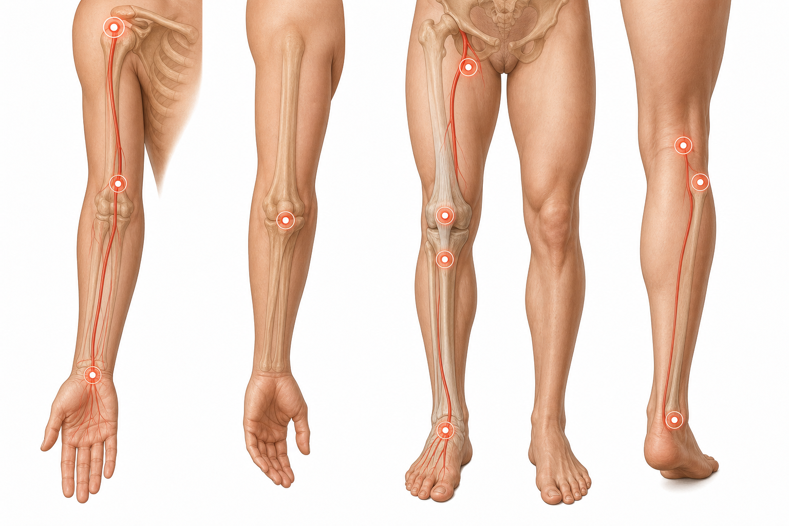
Figure 2. The limb figure complements the torso view by concentrating the pulse-point and landmark regions you would actually palpate during a focused exam.
Surface Markings of Vessels and Nerves
Some structures are not visible but can be estimated from landmarks:
- Radial artery: lateral wrist
- Brachial artery: medial to biceps tendon in the cubital fossa
- Femoral artery: midpoint of the inguinal ligament region
- Sciatic nerve danger zone: gluteal region, avoided by using the superolateral quadrant for intramuscular injection
These surface projections reduce procedural error.
Clinical Applications
Physical Examination
Inspection, palpation, percussion, and auscultation all depend on surface anatomy.
Procedures
- Venipuncture
- Lumbar puncture
- Thoracentesis
- Chest drain insertion
- Intramuscular injection
Emergency Medicine
Rapid recognition of landmarks saves time during trauma and resuscitation.
Key Terms
| Term | Definition | Related Concept |
|---|---|---|
| Surface anatomy | Study of external landmarks related to deeper structures | Physical examination |
| Midclavicular line | Vertical line through midpoint of clavicle | Apex beat, organ projection |
| Sternal angle | Junction of manubrium and sternal body | 2nd rib, intercostal counting |
| Costal margin | Inferior border of rib cage | Liver and upper abdominal exam |
| McBurney's point | Surface point used in appendiceal localization | Right lower quadrant pain |
| Anatomical snuffbox | Triangular depression on dorsolateral wrist | Scaphoid tenderness |
| Safe triangle | Chest wall zone for drain insertion | Pleural procedures |
| Femoral pulse | Palpable arterial pulse below inguinal ligament | Shock, vascular exam |
Common Mistakes
Misconception: Surface anatomy is just about memorizing visible body parts.
Why it's wrong: Surface anatomy is about linking the external body to deeper structures and clinical action.
Correct understanding: Landmarks matter because they help you examine, diagnose, and perform procedures safely.
Misconception: Surface landmarks are identical in every person.
Why it's wrong: Age, obesity, muscle bulk, pregnancy, organ enlargement, and deformity can shift or obscure landmarks.
Correct understanding: Use landmarks as guides, then refine with palpation, percussion, and clinical judgment.
Misconception: If you know the textbook landmark, the procedure is automatically safe.
Why it's wrong: Safe practice also depends on patient position, variation, pathology, and technique.
Correct understanding: Surface anatomy reduces risk, but it does not replace careful procedural reasoning.
Comparison and Connections
| Feature | Bony Landmark | Soft Tissue Landmark | Organ Surface Projection |
|---|---|---|---|
| Reliability | Usually highest | Moderate; varies with tone and body habitus | Variable with respiration and size |
| Example | Iliac crest | Sternocleidomastoid | Liver edge |
| Main use | Orientation | Muscle and vessel localization | Symptom and exam correlation |
| Limitation | Can still vary slightly | Harder in edema or obesity | Less exact than imaging |
Practice Questions
Recall
Q1. Which landmark is used to identify the 2nd rib and count intercostal spaces?
Answer guidance: The sternal angle.
Q2. Which vertebral level is commonly estimated using a line between the iliac crests?
Answer guidance: Around L4 or the L4-L5 interspace.
Understanding
Q3. Why is the anatomical snuffbox clinically important after a fall on the hand?
Answer guidance: Tenderness there suggests a possible scaphoid fracture, which has important blood supply concerns.
Q4. Explain why surface anatomy remains important even when imaging is available.
Answer guidance: Bedside assessment comes first, procedures still require landmarks, and imaging interpretation is easier when clinical localization is strong.
Application
Q5. A clinician needs to give a gluteal intramuscular injection. Which broad region is chosen and why?
Answer guidance: The superolateral gluteal region to reduce risk of sciatic nerve injury.
Q6. A patient has maximal tenderness at McBurney's point. Which diagnosis should be strongly considered?
Answer guidance: Acute appendicitis.
Analysis
Q7. Compare the use of the sternal angle and the midclavicular line in thoracic examination.
Answer guidance: The sternal angle helps count ribs and intercostal spaces; the midclavicular line helps localize structures such as the apex beat and describe findings consistently.
Q8. Why can organ surface projection be less reliable than bony landmarks?
Answer guidance: Organs move with respiration and can change size or position with pathology and body habitus.
FAQ
Q: Why is the sternal angle so frequently mentioned in anatomy and clinical exams?
It is one of the best fixed thoracic landmarks for counting ribs and locating major mediastinal structures.
Q: Can you always feel the liver in a healthy person?
No. A normal liver may not be palpable, especially depending on body habitus and respiratory movement.
Q: Why are pulse points important in surface anatomy?
They help assess circulation, shock, vascular disease, and the location of underlying arteries.
Q: Is the trachea normally exactly in the center?
It is usually midline or nearly so. Noticeable deviation can suggest important thoracic pathology.
Q: Do surface landmarks replace imaging for procedures?
No. They guide bedside practice, but imaging is often used when precision or safety requires it.
Quick Revision
- Surface anatomy links external landmarks to deeper structures.
- Bony landmarks are usually the most reliable reference points.
- The sternal angle marks the 2nd rib and helps count intercostal spaces.
- The apex beat is usually in the 5th left intercostal space near the midclavicular line.
- The iliac crests help estimate the L4-L5 level for lumbar puncture.
- McBurney's point helps localize appendiceal tenderness.
- The anatomical snuffbox is important in suspected scaphoid injury.
- Pulse points and safe procedural zones depend on accurate landmark identification.
- Organ projections are useful but more variable than fixed bony landmarks.
- Surface anatomy is essential for physical examination, bedside diagnosis, and procedures.
Related Topics
Prerequisites: Introduction to Human Anatomy and Osteology.
Related Topics: Gross Anatomy of Thorax, Gross Anatomy of Abdomen, Head and Neck Anatomy.
Next Topics: Use this page alongside the regional gross anatomy chapters so the deeper structures make sense at the bedside.