Head and Neck Anatomy
Learning Objectives
- Identify the major skeletal, muscular, vascular, nervous, and visceral structures of the head and neck.
- Explain how the skull, face, neck triangles, and fascial planes organize this complex region.
- Relate major cranial nerves and cervical structures to their key functions and clinical findings.
- Describe the carotid sheath, airway landmarks, and salivary gland regions in clinically useful terms.
- Apply head and neck anatomy to airway management, stroke localization, thyroid surgery, facial nerve injury, and lymph node assessment.
- Distinguish superficial landmarks from deep structures that carry major procedural risk.
Quick Answer
Head and neck anatomy is one of the most clinically dense regions in medicine. It contains the brain's protective case, the special sense organs, the upper airway, the upper digestive tract, and the major vessels and nerves linking the brain to the rest of the body. Students need this anatomy to understand cranial nerve palsies, facial trauma, thyroid disease, carotid pathology, lymph node spread, airway procedures, and head and neck imaging. The challenge is not just memorizing structures, but seeing how tightly packed and interconnected they are.
Overview
The head and neck region includes:
- The skull and facial skeleton
- The brain and cranial cavity
- Eyes, ears, nose, and oral cavity
- Pharynx and larynx
- Salivary glands
- Cervical fascia and neck compartments
- Major arteries, veins, lymphatics, and nerves
Because so many critical structures pass through a small space, small lesions can produce major symptoms.

Figure 1. This atlas-style overview is most useful for connecting the facial skeleton, salivary region, carotid sheath, larynx, thyroid gland, and cervical vertebrae in one view.
Skull and Facial Skeleton
The skull is divided into:
- Neurocranium: protects the brain
- Viscerocranium: forms the facial skeleton
Key Bones
| Region | Important Bones | Clinical Relevance |
|---|---|---|
| Cranial vault | Frontal, parietal, occipital | Trauma and intracranial protection |
| Skull base | Sphenoid, temporal, ethmoid | Cranial nerve foramina and fracture patterns |
| Face | Maxilla, mandible, zygomatic, nasal bones | Facial trauma and dental relations |
Example
A basilar skull fracture can injure structures passing through foramina and may present with cranial nerve deficits, CSF leak, or periorbital bruising.
Why It Matters
The skull is not a uniform shell. Its foramina, sinuses, and buttresses explain trauma patterns and nerve pathways.
Common Misunderstanding
Students often learn skull bones as isolated names. In clinical anatomy, what matters is what passes through them, what attaches to them, and what breaks near them.
Scalp, Face, and Parotid Region
The scalp has layered anatomy that explains bleeding, infection spread, and surgical dissection.
The face contains:
- Muscles of facial expression
- Branches of the facial nerve
- Trigeminal sensory supply
- Parotid gland and its duct
Parotid Gland
The facial nerve passes through the parotid gland but does not innervate it. This distinction is clinically important in parotid surgery.
Real-World Example
A parotid tumor may present with facial weakness if it involves the facial nerve.
Oral Cavity, Nose, and Pharynx
These structures support breathing, speech, chewing, swallowing, and resonance.
Oral Cavity
- Lips, cheeks, tongue, teeth, palate
- Closely related to salivary glands, muscles, and cranial nerves
Nasal Cavity and Sinuses
- Warm and filter inspired air
- Drain through anatomically specific pathways
Pharynx
- Nasopharynx
- Oropharynx
- Laryngopharynx
Why It Matters
Infections, tumors, and airway obstruction often spread or present according to these compartment boundaries.
Neck Organization
The neck extends from the skull base to the thoracic inlet. It is commonly organized by fascial planes and triangles.
Cervical Fascia
Deep cervical fascia helps compartmentalize structures and can channel the spread of infection.
Anterior and Posterior Triangles
| Triangle | Boundaries and Contents | Clinical Relevance |
|---|---|---|
| Anterior triangle | Sternocleidomastoid, mandible, midline | Carotid arteries, thyroid, larynx, submandibular region |
| Posterior triangle | Sternocleidomastoid, trapezius, clavicle | Accessory nerve, cervical plexus, subclavian region |
Carotid Sheath
Contains:
- Common/internal carotid artery
- Internal jugular vein
- Vagus nerve
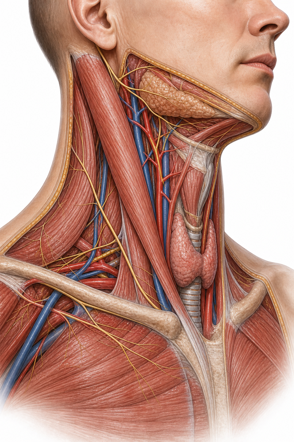
Figure 2. The second plate is especially helpful when revising the sternocleidomastoid, anterior triangle, posterior triangle, thyroid region, and accessory-nerve course.
Real-World Example
Central venous access and carotid surgery depend on precise understanding of carotid sheath relations.
Major Muscles
Key clinically useful muscles include:
- Sternocleidomastoid: rotates and flexes neck; landmark for neck triangles
- Trapezius: shoulder and posterior neck function
- Suprahyoid and infrahyoid groups: swallowing and laryngeal movement
- Muscles of mastication: jaw movement
- Muscles of facial expression: facial movement and emotional display
Blood Supply
The head and neck receive blood primarily from:
- Common carotid arteries
- Internal carotid artery to brain and orbit
- External carotid artery to face, scalp, and neck
- Subclavian arteries
- Vertebral arteries ascending to the brain
Why It Matters
Stroke patterns, epistaxis, scalp bleeding, and surgical control of hemorrhage all depend on vascular anatomy.
Nerve Supply
This region contains major cranial and cervical nerves.
Cranial Nerves with Major Local Relevance
| Nerve | Main Role | Common Clinical Clue |
|---|---|---|
| Trigeminal (V) | Facial sensation, mastication | Facial numbness, weak bite |
| Facial (VII) | Facial expression, taste to anterior tongue | Facial droop |
| Glossopharyngeal (IX) | Taste posterior tongue, swallowing | Loss of gag afferent |
| Vagus (X) | Larynx, pharynx, autonomics | Hoarseness, palatal asymmetry |
| Accessory (XI) | Sternocleidomastoid, trapezius | Shoulder droop |
| Hypoglossal (XII) | Tongue movement | Tongue deviation |
Cervical Plexus and Brachial Relations
The cervical plexus supplies cutaneous regions of the neck and contributes to the phrenic nerve.
Airway and Thyroid Region
Key landmarks:
- Hyoid bone
- Thyroid cartilage
- Cricoid cartilage
- Trachea
- Thyroid gland
Real-World Example
Endotracheal intubation, cricothyrotomy, and thyroid surgery all rely on accurate landmark recognition.
Why It Matters
This is one of the most time-critical anatomy regions in emergency medicine.
Common Misunderstanding
Students often memorize laryngeal cartilages without connecting them to airway access, swallowing, and recurrent laryngeal nerve risk.
Lymphatic Drainage
The head and neck contain rich lymphatic networks, including superficial and deep cervical nodes.
Clinical Relevance
- Infection localization
- Cancer spread and staging
- Neck mass evaluation
Clinical Correlations
Facial Nerve Palsy
Lesions of the facial nerve affect facial expression and can help localize disease.
Thyroid Surgery
The recurrent laryngeal nerve is at risk, and injury may cause hoarseness.
Carotid Disease
Carotid bifurcation disease can affect cerebral perfusion and stroke risk.
Deep Neck Space Infection
Fascial planes can direct spread toward the mediastinum.
Airway Obstruction
Swelling in the tongue, epiglottis, or larynx can become rapidly life-threatening.
Key Terms
| Term | Definition | Related Concept |
|---|---|---|
| Neurocranium | Bony case of the brain | Cranial protection |
| Viscerocranium | Facial skeleton | Facial trauma |
| Carotid sheath | Fascial compartment containing carotid artery, IJV, vagus | Neck surgery |
| Sternocleidomastoid | Major neck muscle and surface landmark | Neck triangles |
| Parotid gland | Major salivary gland in lateral face | Facial nerve relations |
| Recurrent laryngeal nerve | Branch of vagus supplying most intrinsic laryngeal muscles | Hoarseness |
| Trigeminal nerve | Major sensory nerve of face | Facial sensation |
| Facial nerve | Motor nerve to facial expression muscles | Bell palsy |
| Hyoid bone | Suspended bone supporting tongue and larynx | Swallowing |
| Deep cervical fascia | Fascial layers compartmentalizing neck | Infection spread |
Common Mistakes
Misconception: The parotid gland is supplied motor-wise by the facial nerve because the nerve runs through it.
Why it's wrong: The facial nerve traverses the gland but does not provide its parasympathetic secretomotor supply.
Correct understanding: The facial nerve is a surgical relation of the parotid, not its glandular motor supply.
Misconception: Neck triangles are just exam diagrams with little practical value.
Why it's wrong: They help localize vessels, nerves, lymph nodes, glands, and procedural targets.
Correct understanding: Neck triangles are clinical roadmaps.
Misconception: Cranial nerve symptoms can be understood without regional anatomy.
Why it's wrong: Lesion localization depends on where each nerve travels and what nearby structures may affect it.
Correct understanding: Head and neck anatomy is essential for meaningful cranial nerve interpretation.
Comparison and Connections
| Feature | Internal Carotid Artery | External Carotid Artery |
|---|---|---|
| Main territory | Brain and orbit | Face, scalp, much of neck |
| Neck branches | No major branches in neck | Multiple named branches |
| Clinical emphasis | Stroke and intracranial flow | Facial bleeding, surgical anatomy |
| Surface relevance | Deep and less directly palpable | Branch territory explains face/scalp supply |
Practice Questions
Recall
Q1. What three major structures lie in the carotid sheath?
Answer guidance: Carotid artery, internal jugular vein, and vagus nerve.
Q2. Which cranial nerve supplies muscles of facial expression?
Answer guidance: Facial nerve, cranial nerve VII.
Understanding
Q3. Why is the sternocleidomastoid such an important surface landmark?
Answer guidance: It helps define neck triangles and orient major vessels, nerves, and lymphatic structures.
Q4. Explain why deep neck space infections can become dangerous quickly.
Answer guidance: Fascial planes permit spread to other neck compartments and even the mediastinum, threatening airway and systemic stability.
Application
Q5. A patient becomes hoarse after thyroid surgery. Which nerve is classically at risk?
Answer guidance: The recurrent laryngeal nerve.
Q6. A lesion causes weakness in smiling and eye closure on one side of the face. Which nerve is implicated?
Answer guidance: The facial nerve.
Analysis
Q7. Compare the clinical importance of the internal and external carotid arteries in head and neck medicine.
Answer guidance: Internal carotid disease affects intracranial circulation and stroke risk; external carotid branches matter more in facial blood supply and surgery.
Q8. Why does head and neck anatomy produce such diverse symptoms from small lesions?
Answer guidance: The region is densely packed with airway, vascular, neural, sensory, and glandular structures in close proximity.
FAQ
Q: Why is scalp bleeding often dramatic?
The scalp has a rich blood supply and fibrous tissue that holds cut vessels open.
Q: Why are cranial nerve exams so important in head and neck disease?
They help localize lesions to specific foramina, glands, brainstem regions, or neck compartments.
Q: What makes the recurrent laryngeal nerve clinically famous?
Its vulnerability during thyroid and neck surgery and its role in vocal cord movement.
Q: Why do enlarged cervical lymph nodes matter so much?
They may indicate infection, inflammation, or metastatic spread from head and neck malignancy.
Q: Do all facial muscles attach to bone?
No. Many muscles of facial expression insert into skin, which is why they create visible expression.
Quick Revision
- Head and neck anatomy combines airway, sensory organs, vessels, and cranial nerves in a compact region.
- The skull includes neurocranium and viscerocranium.
- Neck triangles are practical guides to local anatomy.
- The carotid sheath contains carotid artery, internal jugular vein, and vagus nerve.
- Facial nerve controls facial expression; trigeminal nerve supplies much facial sensation.
- Parotid gland anatomy is surgically important because the facial nerve passes through it.
- Thyroid and laryngeal landmarks are crucial in airway management and surgery.
- Deep cervical fascia directs infection spread.
- Cervical lymph nodes are central to infection and cancer assessment.
- Small regional lesions can produce major neurological or airway symptoms.
Related Topics
Prerequisites: Introduction to Human Anatomy, Surface Anatomy, Osteology.
Related Topics: Neuroanatomy, Embryology, Otorhinolaryngology, Ophthalmology.
Next Topics: Neuroanatomy to deepen cranial nerve and CNS connections, then later ENT and surgery modules for clinical application.