Gross Anatomy of Abdomen
Learning Objectives
- Describe the boundaries and compartments of the abdomen and explain how they organize abdominal anatomy.
- Identify the major abdominal organs and summarize their positions, peritoneal relationships, and core functions.
- Explain the peritoneum, mesenteries, and omenta in clinically useful terms.
- Trace the major arterial supply, venous drainage, and autonomic innervation of abdominal organs.
- Apply abdominal anatomical knowledge to pain localization, imaging, hernias, appendicitis, and portal hypertension.
- Distinguish intraperitoneal from retroperitoneal structures and explain why that distinction matters clinically.
Quick Answer
The abdomen contains most of the digestive system as well as the spleen, kidneys, major vessels, and complex peritoneal spaces. Gross anatomy of the abdomen matters because pain, bleeding, infection, obstruction, and surgical access all depend on where organs sit and how they relate to each other. A surgeon planning a cholecystectomy, an emergency physician evaluating right lower quadrant pain, and a radiologist reading a CT scan all rely on the same foundational map: abdominal walls, quadrants, peritoneal folds, organ relations, and vascular supply.
Overview of the Abdomen
The abdomen lies between the thorax and pelvis. It is bounded:
- Superiorly by the diaphragm
- Inferiorly by the pelvic inlet and continuity with the pelvis
- Anteriorly and laterally by the abdominal wall
- Posteriorly by the lumbar vertebrae, posterior abdominal wall muscles, and associated fascia
Clinically, the abdomen is often described by four quadrants or nine regions to help localize pain and pathology.
Why It Matters
Localization is not just descriptive. It narrows differentials. Right upper quadrant pain suggests a different set of organs and diseases than left lower quadrant pain.

Figure 1. Use this labeled organ map to orient the liver, stomach, spleen, pancreas, kidneys, bowel loops, and central vessels before moving into peritoneal relationships and clinical correlations.
Abdominal Wall
The abdominal wall protects viscera, supports posture, and generates pressure for coughing, urination, defecation, and childbirth.
Main Layers
- Skin
- Superficial fascia
- Muscular layers
- Transversalis fascia
- Extraperitoneal tissue
- Parietal peritoneum
Major Muscles
| Muscle | Main Action | Clinical Importance |
|---|---|---|
| External oblique | Trunk rotation, compression | Inguinal canal relations |
| Internal oblique | Trunk rotation, compression | Forms part of inguinal wall |
| Transversus abdominis | Compression of abdominal contents | Core stability |
| Rectus abdominis | Trunk flexion | Surface landmark and surgical access |
Real-World Example
Weakness in the lower anterior abdominal wall contributes to inguinal hernia formation.
Common Misunderstanding
Students often picture the abdominal wall as a simple covering. It is actually a multilayered, clinically active structure central to hernias, incisions, and referred pain.
Peritoneum, Mesenteries, and Omenta
The peritoneum is a serous membrane with:
- Parietal peritoneum lining the abdominal wall
- Visceral peritoneum covering organs
Intraperitoneal vs Retroperitoneal
| Category | Organs | Clinical Importance |
|---|---|---|
| Intraperitoneal | Stomach, liver, spleen, jejunum, ileum, transverse colon, sigmoid colon | More mobile; mesenteric relations matter |
| Retroperitoneal | Kidneys, ureters, pancreas except tail, most of duodenum, ascending and descending colon, aorta, IVC | Deep relation to posterior wall and major vessels |
Mesenteries and Omenta
- Mesentery proper: suspends jejunum and ileum.
- Greater omentum: fatty apron from greater curvature of stomach.
- Lesser omentum: connects liver to stomach and proximal duodenum.
Real-World Example
Perforation of a gastric ulcer can spill contents into the peritoneal cavity, producing peritonitis because the peritoneum is highly sensitive and widely distributed.
Why It Matters
Peritoneal relationships determine spread of infection, ascites distribution, and how surgeons reach organs.
Common Misunderstanding
“Retroperitoneal” does not mean outside the abdomen. It means located posterior to the peritoneal lining within the abdominal space.
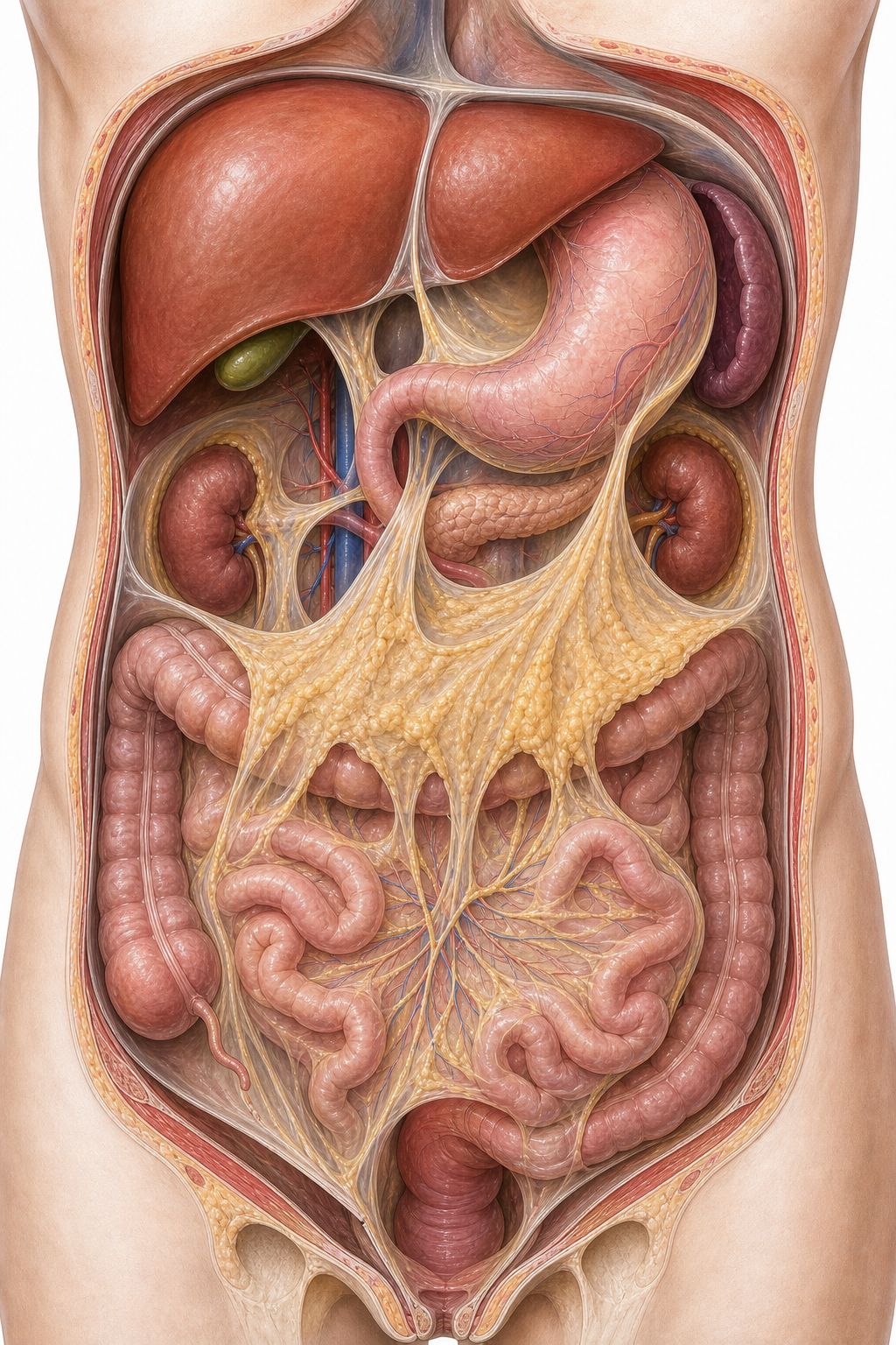
Figure 2. This supplemental plate emphasizes the mesentery, greater omentum, and the posterior position of the kidneys and pancreas compared with most bowel.
Major Abdominal Organs
Liver
The liver lies mainly in the right upper quadrant beneath the diaphragm. It is the largest internal organ and receives dual blood supply from the hepatic artery and portal vein.
Clinical example: Hepatomegaly may push the liver edge below the costal margin.
Gallbladder
Located on the inferior surface of the liver, it stores and concentrates bile.
Clinical example: Gallbladder inflammation often causes right upper quadrant pain and may produce Murphy's sign.
Stomach
The stomach lies mostly in the left upper quadrant between esophagus and duodenum. It has cardia, fundus, body, and pyloric regions.
Clinical example: Posterior gastric ulcers can affect structures behind the stomach, including the pancreas.
Small Intestine
- Duodenum: mostly retroperitoneal and closely related to pancreas
- Jejunum: thicker wall, more vascular
- Ileum: thinner wall, more lymphoid tissue
Clinical example: Small bowel obstruction often presents with colicky pain, vomiting, and dilated loops on imaging.
Large Intestine
Includes cecum, appendix, colon, rectum, and anal canal. Its position helps localize common disorders like appendicitis or sigmoid volvulus.
Pancreas
A mostly retroperitoneal gland extending from the duodenum toward the spleen.
Clinical example: Pancreatitis pain often radiates to the back because of posterior relations.
Spleen
Located in the left upper quadrant deep to ribs 9 to 11. It is clinically important in trauma because enlargement or rupture can cause serious hemorrhage.
Kidneys and Ureters
The kidneys are retroperitoneal on the posterior abdominal wall. Pain from ureteric stones often radiates from flank to groin.
Blood Supply and Drainage
Arterial Supply
The abdominal aorta gives three major unpaired anterior branches to the gut:
- Celiac trunk: foregut
- Superior mesenteric artery: midgut
- Inferior mesenteric artery: hindgut
Venous Drainage
Most abdominal gastrointestinal drainage enters the portal venous system, which carries nutrient-rich blood to the liver before reaching systemic circulation.
Why It Matters
Vascular territories explain ischemia patterns, surgical ligation strategies, and portal-systemic complications.
Real-World Example
Portal hypertension can produce varices because venous blood is forced through collateral pathways.
Nerve Supply
Abdominal organs receive autonomic innervation:
- Parasympathetic: mainly via vagus nerve to foregut and midgut; pelvic splanchnics for hindgut
- Sympathetic: via thoracic and lumbar splanchnic pathways
Visceral afferents travel with autonomics and help explain referred pain patterns.
Example
Early appendicitis often causes vague periumbilical pain because visceral afferents from the midgut refer pain to the T10 region before parietal peritoneum becomes involved.
Important Spaces and Recesses
Abdominal spaces matter because fluid, blood, or infection collects in predictable places.
- Subphrenic spaces
- Hepatorenal recess (Morison pouch)
- Lesser sac
- Paracolic gutters
Real-World Example
In a supine patient with intra-abdominal fluid, Morison pouch is a common collection site and is checked during FAST ultrasound.
Clinical Correlations
Appendicitis
Pain often migrates from periumbilical to right lower quadrant as inflammation spreads from visceral to parietal peritoneum.
Hernias
Inguinal hernias follow abdominal wall weak points and are closely tied to abdominal wall anatomy.
Portal Hypertension
Raises pressure in the portal system, contributing to varices, ascites, and splenomegaly.
Peritonitis
Inflammation of the peritoneum causes guarding, rebound tenderness, and severe pain.
Abdominal Trauma
Solid organs such as liver and spleen bleed; hollow viscus injury risks contamination and peritonitis.
Key Terms
| Term | Definition | Related Concept |
|---|---|---|
| Peritoneum | Serous membrane lining abdominal cavity and covering organs | Peritonitis |
| Mesentery | Double layer of peritoneum suspending bowel | Vascular supply |
| Greater omentum | Fatty peritoneal fold hanging from stomach | Infection containment |
| Retroperitoneal | Posterior to the peritoneum | Kidneys, pancreas |
| Celiac trunk | Arterial supply to foregut | Upper abdominal organs |
| Portal vein | Venous channel draining GI tract to liver | Portal hypertension |
| Morison pouch | Hepatorenal recess | FAST scan |
| McBurney's point | Surface landmark in appendiceal region | Appendicitis |
| Foregut | Embryologic GI segment supplied by celiac trunk | Stomach, liver, spleen relations |
| Midgut | Segment supplied by SMA | Jejunum, ileum, appendix |
Common Mistakes
Misconception: The abdomen is just a container full of loosely arranged organs.
Why it's wrong: Organ position, peritoneal relations, vascular territories, and spaces are highly organized and clinically meaningful.
Correct understanding: The abdomen is a structured anatomical region where location and relation often determine disease presentation.
Misconception: Pain location always identifies the exact organ involved.
Why it's wrong: Visceral pain is often vague and referred; exact localization often improves only when parietal peritoneum becomes involved.
Correct understanding: Abdominal pain patterns reflect both organ innervation and peritoneal involvement.
Misconception: Retroperitoneal organs are separate from abdominal pathology.
Why it's wrong: Retroperitoneal structures are central to abdominal imaging, trauma, pancreatitis, vascular disease, and posterior pain patterns.
Correct understanding: Retroperitoneal anatomy is essential for understanding deep abdominal disease.
Comparison and Connections
| Feature | Intraperitoneal Organ | Retroperitoneal Organ |
|---|---|---|
| Covering | More completely invested by visceral peritoneum | Posterior to peritoneum |
| Mobility | Usually greater | Usually less |
| Example | Stomach | Kidney |
| Surgical implication | Often accessed by mobilizing peritoneal folds | Often approached via posterior or deep dissection |
| Pain pattern | Often influenced by visceral peritoneum | May present with deep, back-related pain |
Practice Questions
Recall
Q1. Name the three major unpaired branches of the abdominal aorta that supply the gut.
Answer guidance: Celiac trunk, superior mesenteric artery, and inferior mesenteric artery.
Q2. Which abdominal recess is commonly assessed for free fluid on FAST examination?
Answer guidance: Morison pouch, the hepatorenal recess.
Understanding
Q3. Why does early appendicitis often cause periumbilical pain rather than immediate right lower quadrant pain?
Answer guidance: Early pain is visceral and referred from the midgut; later localization occurs when parietal peritoneum is irritated.
Q4. Explain why intraperitoneal and retroperitoneal distinctions matter in surgery.
Answer guidance: They affect organ mobility, exposure, fluid spread, and access routes.
Application
Q5. A patient with portal hypertension develops esophageal varices. What anatomical system is primarily responsible?
Answer guidance: Elevated pressure in the portal venous system with diversion through portosystemic anastomoses.
Q6. A patient has severe left upper quadrant pain after trauma. Which organ is especially concerning?
Answer guidance: The spleen, because of its location and risk of hemorrhage.
Analysis
Q7. Compare why pancreatitis pain and appendicitis pain are felt differently.
Answer guidance: Pancreatic pain often feels deep and may radiate to the back due to retroperitoneal relations; appendiceal pain often evolves from vague visceral pain to localized parietal pain.
Q8. Why can knowledge of abdominal spaces change the interpretation of imaging?
Answer guidance: Fluid, pus, air, or blood spread along predictable recesses and gutters, helping localize the source and severity of disease.
FAQ
Q: Why are abdominal quadrants useful if organs overlap multiple regions?
They simplify bedside localization and communication, even though exact anatomy is more complex.
Q: Is the pancreas completely retroperitoneal?
Most of it is, but the tail is closely related to the spleen and is more mobile in relation to peritoneal structures.
Q: Why is the portal vein so important?
It channels venous blood from much of the GI tract to the liver, making it central to metabolism and portal hypertension.
Q: What is the easiest way to remember foregut, midgut, and hindgut supply?
Link them to arteries: celiac trunk, SMA, and IMA.
Q: Why does diaphragmatic irritation sometimes cause shoulder pain?
Because the diaphragm is innervated by the phrenic nerve, and irritation can refer pain to the C3-C5 shoulder region.
Quick Revision
- The abdomen lies between thorax and pelvis and is bounded superiorly by the diaphragm.
- The abdominal wall is layered and clinically important in hernias and surgical access.
- Peritoneum has parietal and visceral layers.
- Intraperitoneal organs are generally more mobile than retroperitoneal organs.
- Major gut arteries are the celiac trunk, SMA, and IMA.
- Portal venous drainage is central to liver function and portal hypertension.
- The liver, stomach, intestines, pancreas, spleen, kidneys, and gallbladder have distinct positional relationships.
- Morison pouch and paracolic gutters help predict fluid collection.
- Referred abdominal pain reflects autonomic innervation and embryologic gut divisions.
- Gross abdominal anatomy is essential for surgery, emergency medicine, and imaging interpretation.
Related Topics
Prerequisites: Introduction to Human Anatomy, Surface Anatomy, and Thorax anatomy.
Related Topics: Embryology, Histology, General Surgery, Gastrointestinal Physiology.
Next Topics: Head and Neck Anatomy or later Physiology and Pathology chapters to connect abdominal structure with function and disease.