Embryology
Learning Objectives
- Define embryology and explain why developmental anatomy is essential for clinical reasoning.
- Describe the sequence from fertilization through implantation, gastrulation, folding, and organogenesis.
- Identify the three germ layers and summarize the major tissues derived from each.
- Explain basic neurulation, cardiovascular development, and limb development in broad clinical terms.
- Apply embryological reasoning to congenital anomalies, ectopic pregnancy, neural tube defects, and gut malrotation.
- Distinguish embryonic stages by timing and major developmental events.
Quick Answer
Embryology is the study of how the human body develops from fertilization to fetal life. It matters because many adult structures make sense only when you know how they formed. Congenital heart disease, neural tube defects, cleft palate, malrotation, Meckel diverticulum, and undescended testes are all easier to understand when you know the normal developmental pathway. For medical students, embryology is not just a timeline to memorize. It is the logic behind why structures end up where they are, why anomalies cluster in certain patterns, and why some diseases present the way they do.
What Is Embryology?
Embryology studies the formation of the embryo and the earliest stages of fetal development. It links anatomy, genetics, histology, and pathology.
The subject is often challenging because many events happen quickly, but the major idea is simple: the mature body is built through ordered steps, and when one step fails, a recognizable anomaly can result.
Major Developmental Timeline
| Stage | Approximate Timing | Main Event |
|---|---|---|
| Fertilization | Day 0 | Sperm and oocyte fuse to form zygote |
| Cleavage | Days 1-3 | Rapid mitotic divisions without growth |
| Morula | Days 3-4 | Solid ball of cells |
| Blastocyst | Days 5-6 | Inner cell mass and trophoblast form |
| Implantation | Days 6-10 | Blastocyst embeds in endometrium |
| Gastrulation | Week 3 | Three germ layers form |
| Neurulation and folding | Weeks 3-4 | Neural tube and body plan develop |
| Organogenesis | Weeks 4-8 | Major organs begin forming |

Figure 1. This sequence is designed for timeline revision: fertilization, zygote formation, cleavage, morula, blastocyst, uterine-tube transport, and implantation.
Fertilization, Cleavage, and Implantation
Fertilization
Fertilization usually occurs in the uterine tube, creating a zygote with a full diploid chromosome set.
Cleavage
The zygote divides repeatedly without overall enlargement, producing progressively smaller blastomeres.
Blastocyst
The blastocyst has:
- Inner cell mass: forms the embryo proper
- Trophoblast: contributes to placenta
Implantation
The blastocyst implants into the endometrium. Proper implantation is essential for normal pregnancy.
Real-World Example
Implantation outside the uterine cavity causes ectopic pregnancy, which can become life-threatening if the tube ruptures.
Why It Matters
The earliest stages explain infertility, miscarriage, ectopic pregnancy, and early pregnancy testing.
Common Misunderstanding
Students often think implantation happens immediately after fertilization. In reality, the embryo travels and changes form first.
Gastrulation and Germ Layers
Gastrulation forms the three primary germ layers:
- Ectoderm
- Mesoderm
- Endoderm
Major Derivatives
| Germ Layer | Key Derivatives | Clinical Relevance |
|---|---|---|
| Ectoderm | Epidermis, nervous system, neural crest derivatives | Neural tube defects, pigment disorders |
| Mesoderm | Muscle, bone, connective tissue, cardiovascular system, kidneys | Congenital heart and urogenital anomalies |
| Endoderm | Epithelial lining of GI and respiratory tracts and related glands | GI and respiratory developmental defects |
Why It Matters
Most congenital anomalies can be understood better when you know which layer produced the affected tissue.
Common Misunderstanding
Germ layers are not abstract lists for memorization. They are the developmental source code of organ systems.
Embryonic Folding and Body Plan
Folding transforms a flat trilaminar disc into a three-dimensional body form. This process creates:
- Ventral body wall organization
- Gut tube formation
- Relative positioning of heart and foregut
- Early umbilical connection patterns
Real-World Example
Failure of proper ventral wall formation contributes to defects such as omphalocele and gastroschisis.
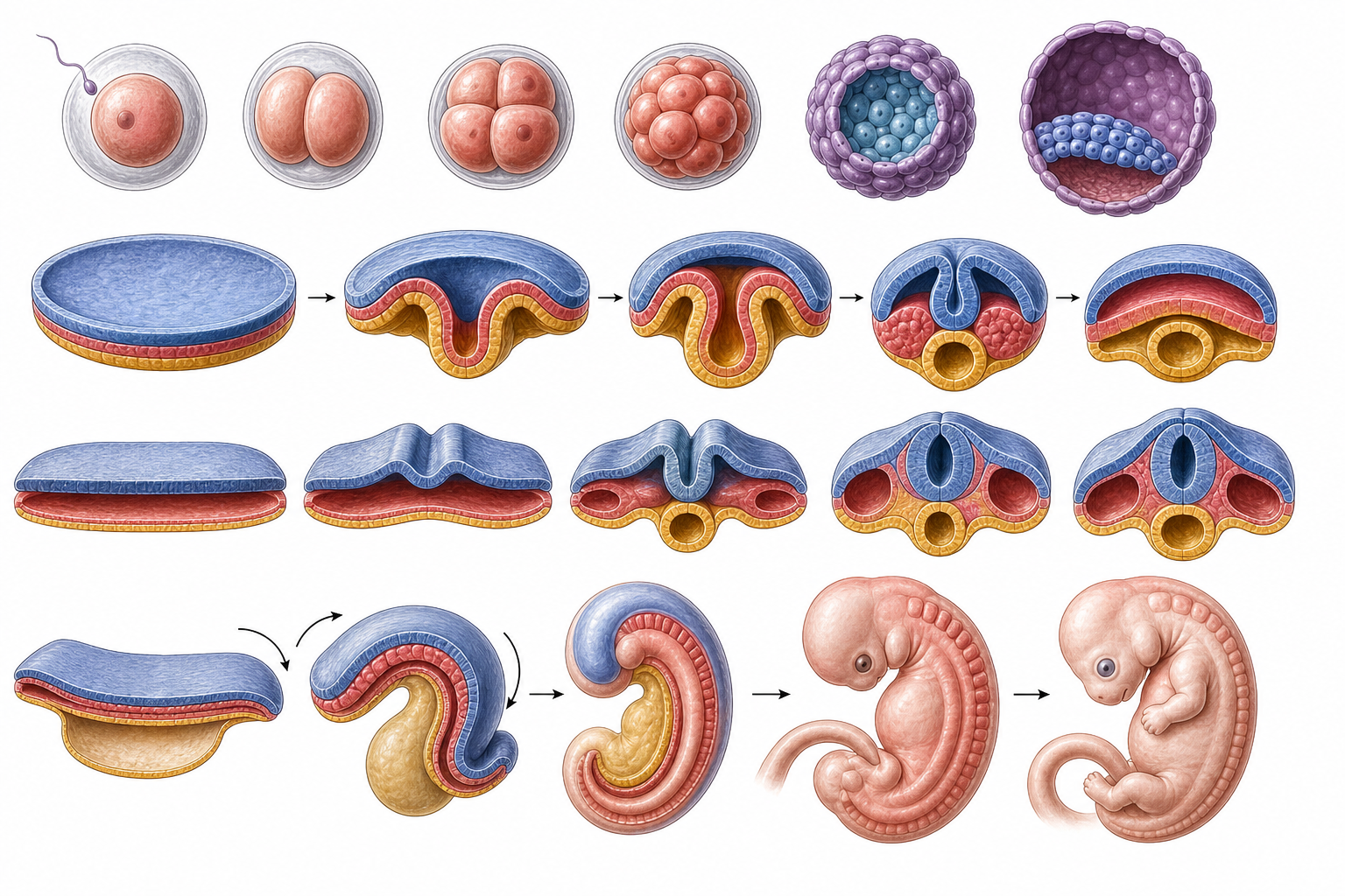
Figure 2. The second plate works best alongside the germ-layer and folding sections because it shows how a flat embryonic disc becomes a three-dimensional body plan.
Neurulation
The neural plate folds to form the neural tube, which gives rise to the central nervous system.
Clinical Relevance
Failure of tube closure can cause:
- Anencephaly
- Spina bifida
Adequate folate intake reduces risk of neural tube defects.
Cardiovascular Development
The heart is one of the earliest functioning organs in the embryo.
Major concepts:
- Fusion of primitive heart tubes
- Cardiac looping
- Septation into chambers
- Remodeling of fetal circulation
Real-World Example
Septation defects help explain congenital heart diseases such as atrial or ventricular septal defects.
GI and Respiratory Development
The primitive gut tube develops into foregut, midgut, and hindgut structures. Budding and partitioning also create respiratory structures.
Clinically Important Concepts
- Midgut rotation
- Vitelline duct remnants
- Tracheoesophageal separation
Example
Failure of normal midgut rotation can lead to malrotation and volvulus.
Limb and Face Development
Limb buds appear early and grow through coordinated signaling and patterning.
Face formation requires fusion of multiple prominences. Disturbances in this process can cause cleft lip or cleft palate.
Why It Matters
Developmental timing helps explain why some anomalies occur together.
Placenta and Fetal Circulation
The placenta supports exchange of gases, nutrients, and wastes between maternal and fetal circulations.
Important fetal circulation features include:
- Ductus venosus
- Foramen ovale
- Ductus arteriosus
These structures normally change after birth.
Clinical Correlations
Ectopic Pregnancy
Implantation outside the uterine cavity, most commonly in the fallopian tube.
Neural Tube Defects
Failure of proper closure during early neurulation.
Congenital Heart Disease
Can result from abnormal septation, outflow tract development, or neural crest contributions.
Meckel Diverticulum
Persistence of the vitelline duct remnant.
Malrotation
Abnormal intestinal rotation can predispose to volvulus and obstruction.
Key Terms
| Term | Definition | Related Concept |
|---|---|---|
| Zygote | Fertilized egg cell | Cleavage |
| Blastocyst | Early embryonic stage with inner cell mass and trophoblast | Implantation |
| Trophoblast | Outer layer of blastocyst contributing to placenta | Pregnancy support |
| Gastrulation | Formation of three germ layers | Organ system origins |
| Ectoderm | Outer germ layer | Nervous system, epidermis |
| Mesoderm | Middle germ layer | Muscle, bone, cardiovascular system |
| Endoderm | Inner germ layer | Gut and respiratory epithelium |
| Neurulation | Formation of neural tube | CNS development |
| Organogenesis | Formation of organs | Embryonic weeks 4-8 |
| Neural crest | Specialized embryonic cell population with diverse derivatives | Face, peripheral nerves, conotruncal heart development |
| Ductus arteriosus | Fetal shunt between pulmonary artery and aorta | Postnatal closure |
| Vitelline duct | Embryonic connection to yolk sac | Meckel diverticulum |
Common Mistakes
Misconception: Embryology only matters for pediatrics and board exams.
Why it's wrong: Developmental anatomy explains adult relationships, congenital disease, and many surgical and radiological findings.
Correct understanding: Embryology helps clinicians reason about both pediatric and adult anatomy.
Misconception: Congenital anomalies are random isolated defects.
Why it's wrong: Many anomalies reflect disruption of specific developmental processes at predictable times.
Correct understanding: Timing and mechanism are central to embryological diagnosis.
Misconception: Germ layers are enough to explain everything on their own.
Why it's wrong: Germ layers are foundational, but folding, migration, induction, vascular development, and fusion events are also critical.
Correct understanding: Embryology is a sequence of interacting processes, not just three derivative lists.
Comparison and Connections
| Feature | Ectoderm | Mesoderm | Endoderm |
|---|---|---|---|
| Broad role | Outer covering and nervous system | Structural and connective systems | Epithelial lining of internal tubes |
| Classic derivative | Brain and epidermis | Muscle and bone | Intestinal epithelium |
| Common anomaly link | Neural tube defects | Cardiac and renal anomalies | GI and airway partition defects |
Practice Questions
Recall
Q1. What are the three primary germ layers formed during gastrulation?
Answer guidance: Ectoderm, mesoderm, and endoderm.
Q2. Where does fertilization most commonly occur?
Answer guidance: In the uterine tube, usually the ampullary region.
Understanding
Q3. Why is the blastocyst stage important for implantation?
Answer guidance: It contains the trophoblast for implantation and the inner cell mass for embryo formation.
Q4. Explain why neurulation defects can have severe consequences.
Answer guidance: The neural tube gives rise to the brain and spinal cord, so failed closure affects core CNS development.
Application
Q5. A newborn has a midline lower back defect with neural tissue involvement. Which developmental process most likely failed?
Answer guidance: Proper neural tube closure during neurulation.
Q6. A child develops intestinal obstruction due to abnormal gut rotation. Which broad embryological process is implicated?
Answer guidance: Midgut rotation during embryonic development.
Analysis
Q7. Compare ectopic pregnancy and failed implantation in terms of developmental stage.
Answer guidance: Both involve the implantation phase, but ectopic pregnancy reflects implantation in the wrong location rather than failure of implantation altogether.
Q8. Why does embryology help explain adult anatomical oddities such as recurrent laryngeal nerve pathways or gut rotation?
Answer guidance: Adult anatomy reflects developmental movement, migration, and remodeling rather than simple static design.
FAQ
Q: Why do medical students find embryology difficult at first?
Because it is highly dynamic and time-based. It becomes easier when learned as a sequence of transformations rather than isolated facts.
Q: What is the most high-yield embryology skill for exams and clinics?
Connecting a defect to the developmental step that failed.
Q: Why is folate important in pregnancy?
Adequate folate reduces the risk of neural tube defects during early development.
Q: Does organogenesis continue throughout pregnancy?
Major organ formation is concentrated in the embryonic period, while later fetal development focuses more on growth and maturation.
Q: Why is timing so important in teratology?
Different organs are vulnerable at different developmental windows, so timing shapes the type of anomaly produced.
Quick Revision
- Embryology explains how normal anatomy forms and why anomalies occur.
- Fertilization is followed by cleavage, morula, blastocyst formation, and implantation.
- Gastrulation forms ectoderm, mesoderm, and endoderm.
- Folding converts the flat disc into a three-dimensional body plan.
- Neurulation forms the neural tube.
- Organogenesis is most active during weeks 4 to 8.
- The heart develops early and requires looping and septation.
- Gut rotation and body wall closure are major developmental events.
- Congenital anomalies often reflect failure of a specific developmental step.
- Embryology connects anatomy, pathology, pediatrics, surgery, and radiology.
Related Topics
Prerequisites: Introduction to Human Anatomy and basic cell biology.
Related Topics: Histology, Neuroanatomy, Obstetrics and Gynecology, Pediatrics, Pathology.
Next Topics: Use embryology alongside regional anatomy to understand why adult structures have their final arrangement.